Brain – The Atlas of the Human Brain in Stereotaxic Space
A short introduction in to the Atlas of the Human Brain and the Brain used throughout for the research on this site. To take a more systematical approach to the use of the provided material both on the DVD from the "Atlas of the Human Brain" and the applications you can find on this website the following explanatory steps might help.
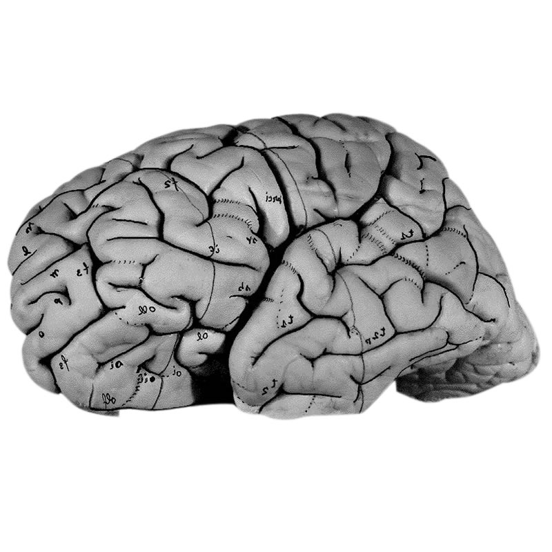
Figure 1:
The Brain used for research in the "Atlas of the Human Brain" and which are used for the main
applications is from a 24-year-old male from the Vogt collection in Düsseldorf.
Further details about The Brain.
The overall layout of the Atlas and the website is based on the diffentiation of the macroscopic area which is mainly represented in the Head & Brain area.

Figure 2:
After determining the surface of the brain the brain is cut in 5 blocks prior to the sectioning process according to the sterotaxic space. Photographs and diagrams are available in the "Atlas of the Human Brain" and in the sections area whereas you can also find the very brain in the corresponding virtual microscopy (nissl staining).

Figure 3:
Following the delineation process based on analysis of the cyto- and myelostructure of each slice, there are several reconstructions in three dimensions. The 3D Models of the thalamic and subthalamic structures, distinguished nuclei and their subdivisions are visually represented in the 3D Reconstruction area.
Material and applications in this section
Surface Views
 View of the external morphology of the formalin fixed atlas brain before slicing in different angles as presented in the Atlas of the Human Bain.
Sections
 The sectional Brain Atlas focuses only on the anatomical structures while both the schematic and slice view are used to represent the area of interest.
Virtual Microscopy
![]() The nissl stained Atlas Brain has been prepared for the virtual microscopy to explore the anatomical structures of the human brain at the cell level.
The nissl stained Atlas Brain has been prepared for the virtual microscopy to explore the anatomical structures of the human brain at the cell level.
AHB-Literature
 Here you may retrieve relevant descriptive and quantitative data, including morphometric data
Literature
Structures
Numerous desciptive and quantitative studies have been performed on the brain represented in the Myeloarchitectonic Atlas.
To the structures
BrainSlicer
 BrainSlicer allows to interactively navigate within the 3D atlas brain in real time to visualize segmented cortical and ...
3D Views
 3D (three-dimensional) reconstructions "Contour-lines" defining pial and ventricular surfaces, borderlines around ...
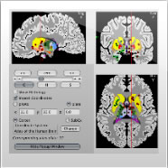
Transformation
 Interpretations of CT or MRI dataset by the integration with the resource provided by the Atlas of the Human Brain are computed to obtain precise anatomical orientation.
BrainNavigator
 The BrainNavigator allows 3D navigation through the detailed atlas of the isolated brain.
Locator
 The program shows the orthogonal planes to which these coordinates are common. With this tool it is possible to dial ...

